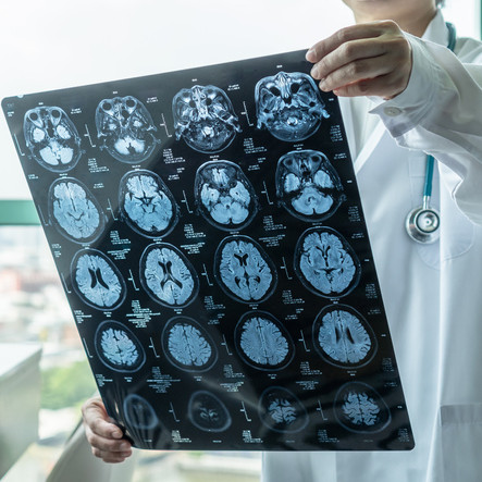
Traumatic brain injuries claim over 166 lives daily and are usually characterized by nausea, headache, drowsiness, mood swings, clumsiness, and lack of coordination.
It may be tempting to brush off mild headaches after a car accident, especially if one looks outwardly fine. However, did you know headaches can result from a traumatic brain injury suffered during a car accident?
Traumatic brain injuries (TBIs) claim over 166 lives every day and are usually characterized by nausea, headache, drowsiness, mood swings, clumsiness, and lack of coordination. Let’s consider some common medical tests used in TBI diagnosis.
The Tests
Glasgow Coma Scale
The GCS rapidly assesses mental operations using pupil measurements, eye movements, touch response, physical reflexes, speech ability, and obedience to verbal commands.
After the assessment, the doctor will allocate a GCS reading. Generally, a 3-rating usually indicates a comma, and a 15-rating indicates normal function. Anything in between may indicate mild or moderate brain injuries.
CT or CAT
Computerized Tomography uses powerful X-rays to capture clear brain images in many thin sheets and then combines them to form a whole-brain image. They are very effective in diagnosing intracranial hemorrhages, bruised brain tissues, skull fractures, and other forms of brain damage. Sometimes, the technician may inject you with a radioactive dye to enhance the diagnosis.
Magnetic Resonance Imaging
You’ve probably seen those tunnel-like machines in movies where the automatic bed moves the patient’s head inside the equipment. That is an MRI. The machine operates similarly to a CT scan, only that it uses powerful magnets in place of X-rays. Doctors generally conduct MRIs for emergency diagnosis when the patient shows no improvement or during follow-up examination.
Electroencephalogram
EEG tests supplement MRI when testing for brain activity and injuries.
Brain activity produces electric currents, which the equipment detects using small electrical conductors attached to your scalp. Abnormal electrical signals may indicate the presence of a TBI.
Positron Emission Tomography Scan
A PET scan allows doctors to observe tissue and organ operation. The doctor typically injects the patient with a small amount of a radioactive substance called a tracer. And then use special equipment to trace its path inside the body. High chemical activity around specific organs or tissues is usually an indication of damage or disease.
Diffusion Tensor Imaging (DTI)
This is an advanced form of MRI because it uses magnetic resonance to create brain images. The only difference between the two is that it can also track the diffusion of water molecules within the white matter fibers that run between different brain sections. Abnormal water diffusion in those pathways could signify TBIs within the brain’s microscopic structure.
St. Louis Car Accident Attorneys
Have you or a loved one recently been involved in a car accident that resulted in severe brain injuries? The experienced St. Louis car accident attorneys at The Hoffman Law Firm can help evaluate your claim for free and help you determine what compensation you may be entitled to for your medical costs, lost wages, and other injury-related expenses. Give us a call 24/7 for a free consultation.
FREE Consultation
Speak with an experienced attorney 24/7

